Upper Leg Tendon Anatomy / Muscles Of The Anterior Thigh Quadriceps Teachmeanatomy : The patella is attached to the shinbone (tibia) by the patellar tendon.
Upper Leg Tendon Anatomy / Muscles Of The Anterior Thigh Quadriceps Teachmeanatomy : The patella is attached to the shinbone (tibia) by the patellar tendon.. Upper leg tendon anatomy : It flexes the thigh at the hip joint, and extends at the knee joint. The patella is attached to the shinbone (tibia) by the patellar tendon. On the medial edge of the posterior thigh is the gracilis muscle. The thigh has some of the body's largest muscles.
Other muscles of the anterior (front) thigh include the pectineus, sartorius, and the. Notice the upper leg has a biceps muscle just like the upper arm does. The thigh muscles are divided into three compartments: This is why you have to indicate which biceps you are taking about when discussing one or other of these muscles. Collectively, the muscles in this area plantarflex and invert the foot.
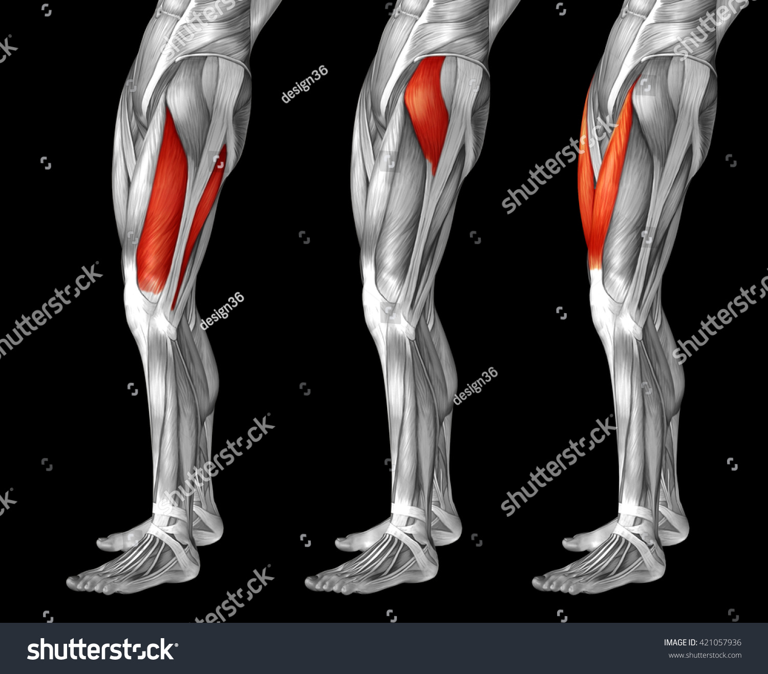
Concept 3d Human Upper Leg Anatomy Stock Illustration 421057936 from image.shutterstock.com Anatomy the four quadriceps muscles meet just above the kneecap (patella) to form the quadriceps tendon. These muscles run from the lower spine. It is also visible on the medial edge of the thigh from the anterior. One of the most important tendons in terms of mobility of the leg is the achilles tendon. Collectively, the muscles in this area plantarflex and invert the foot. These four muscles come together to form a single tendon, which inserts into the patella, or kneecap. The largest muscle masses in the leg are present in the thigh and the calf. Related posts of muscle anatomy of upper thigh muscle anatomy crossword key biology corner.
Upper leg tendon anatomy / anatomical structures and specific regions are visible as dynamic labeled images.
Squeeze your knees together and boom, you're contracting the adductors. Case contributed by dr roberto schubert. Your hamstring muscles connect to the back of your knee via the hamstring tendon. Legs are used for standing, and all forms of. It's the area that runs from the hip to the knee in each leg. These muscles run from the lower spine. The detailing of these structures changes based on dog breed due to the huge variation of size in dog breeds. Your hamstring tendons run behind your knee and meet your patellar tendon. It runs straight down the leg and attaches to the patella via the quadriceps femoris tendon. A muscle strain is graded progressively determined by the number of muscle or tendon fibers that tear during injury. 1 article features images from this case. Collectively, the muscles in this area plantarflex and invert the foot. The posterior upper leg muscles provide your knees with mobility (extension, flexion and rotation) and strength.they work closely with your quadriceps muscles at the front of your thigh, your gluteal muscles, and your calf muscles to ensure proper movement of your leg and hip.
Your hamstring muscles connect to the back of your knee via the hamstring tendon. Muscles of the lower limb; These muscles run from the lower spine. Rectus femoris muscle, one of the quadriceps muscles on the front of your thigh. Collectively, the muscles in this area plantarflex and invert the foot.
Anatomy Of The Hamstring Muscles from www.verywellfit.com Other muscles of the anterior (front) thigh include the pectineus, sartorius, and the. Collectively, the muscles in this area plantarflex and invert the foot. One of the most important tendons in terms of mobility of the leg is the achilles tendon. Upper leg anatomy and function the upper leg is often called the thigh. The patella is attached to the shinbone (tibia) by the patellar tendon. Muscle anatomy crossword key biology corner 12 photos of the muscle anatomy crossword key biology corner muscle anatomy crossword answer key biology corner, muscle anatomy crossword key biology corner, muscle anatomy crossword puzzle answers biology corner, human muscles, muscle anatomy crossword. In clinical anatomy the thigh muscles are divided into three groups: Leg muscles anatomy leg anatomy muscle anatomy thigh muscles.
Protecting, resting, applying ice, compression (such as wrapping the area with an elastic bandage), and elevation are good treatments for most calf muscle.
The quadriceps tendon attaches the quadriceps muscles to the patella. Grade 1 is a mild strain in which a small number of fibers tear. Muscle anatomy crossword key biology corner 12 photos of the muscle anatomy crossword key biology corner muscle anatomy crossword answer key biology corner, muscle anatomy crossword key biology corner, muscle anatomy crossword puzzle answers biology corner, human muscles, muscle anatomy crossword. We created an anatomical atlas of the upper limb, an. On the medial edge of the posterior thigh is the gracilis muscle. It's the area that runs from the hip to the knee in each leg. Upper leg, knee, lower leg, ankle, and foot. People who play soccer have these specific muscles of the leg very well defined, so they're like a walking anatomy atlas for thigh muscles. Grade 1 strains can produce a sharp pain, but it is usually a feeling of tightness or a pulling sensation. The thigh has some of the body's largest muscles. Medial muscles adduct and rotate your thigh, and posterior flex your leg and extend your thigh. Diagnosis not applicable diagnosis not applicable. Dog anatomy details the various structures of canines (e.g.
Case contributed by dr roberto schubert. Upper leg tendon anatomy / biology 2404 a&p basics. Leg muscles anatomy leg anatomy muscle anatomy thigh muscles. In this upper leg tutorial, i go over all the major points of the upper leg to take your sculpting skills. Learn about our editorial process.
The Muscles Of The Upper Legs Rear Spc Id 2137 Science 3d Illustration from www.sciencepicture.co Upper leg anatomy and function the upper leg is often called the thigh. The detailing of these structures changes based on dog breed due to the huge variation of size in dog breeds. We created an anatomical atlas of the upper limb, an. People who play soccer have these specific muscles of the leg very well defined, so they're like a walking anatomy atlas for thigh muscles. The thigh is the region between the hip and knee joints. Leg muscles anatomy leg anatomy muscle anatomy thigh muscles. The thigh muscles are divided into three compartments: This is the physiological upper limit of tendon strain whereby the collagen fibrils orient themselves in the direction of tensile mechanical.
Upper leg tendon anatomy :
This chart is beautifully illustrated and offers the most comprehensive look at the muscles of the human leg available. The patella is attached to the shinbone (tibia) by the patellar tendon. The thigh has some of the body's largest muscles. It is the junction of the thigh and the leg and is a hinge joint. The anatomy of the iliacus muscle hip flexor that flexes and rotates the thigh bone. On the medial edge of the posterior thigh is the gracilis muscle. Notice the upper leg has a biceps muscle just like the upper arm does. Thigh muscles are responsible for allowing normal gait and proper lower extremity function (1). Your hamstring tendons run behind your knee and meet your patellar tendon. In clinical anatomy the thigh muscles are divided into three groups: Anatomy the four quadriceps muscles meet just above the kneecap (patella) to form the quadriceps tendon. The medial thigh muscles are responsible for the adduction (movement of a body part toward the body's midline) of the leg. Diagnosis not applicable diagnosis not applicable.
:max_bytes(150000):strip_icc()/GettyImages-87308179-56a05f563df78cafdaa14cd4.jpg)
